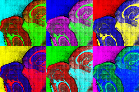
A chemical map of cancer tissue

Researchers from the University of Gothenburg are using a special mass spectrometry method — normally deployed in the analysis of computer chips, lacquers and metals — to better detect harmful cells in the body.
As part of her doctoral thesis, Tina Angerer sought to study the distribution of lipids — the main building blocks of cell membranes — in cancer tissue. Lipids can change rapidly and reveal problems in cells; for this reason, they are one of several keys to understanding cancer.
Angerer’s method of choice was imaging time-of-flight secondary ion mass spectrometry (ToF-SIMS), which works by shooting gas projectiles at a substance, releasing molecules and atoms from that substance in the process. Mass spectrometry is used to identify these particles, and the process is repeated until the entire surface has been analysed.
But while ToF-SIMS has high enough sensitivity to detect lipid distributions on a subcellular scale in biological samples, there is one drawback: it is a highly destructive process which typically destroys a lot of the information it acquires. For this reason, it is typically used to analyse inorganic materials such as computer chips, lacquers, rust, semiconductors and metals.
Angerer and her colleagues solved this problem by combining gas cluster ion beams (GCIBs) with trifluoracetic acid (TFA) vapour exposure, optimising the method for organic and biological materials in the process. “With our instruments,” she said, “the ‘bombardment’ is softer, so that large molecules can be analysed at the same time as the chemical pictures we take and still have good resolution.”
Deploying the method in cancer tissue, the researchers found that while most cells in the body build their lipids with fatty acids derived from the food we eat, cancer cells build their fatty acids by using various types of sugar, as well as fat and protein. Scientists have long known that cancer cells consume great amounts of sugar and have suggested that they may create their own fatty acids, but there was no evidence of such a thing until now.
“We have been able to show that fatty acids from food can be found around cancer tissue and in healthy cells, but that cancer cells consist almost exclusively of fatty acids that the cells have created by themselves,” said Angerer. “This means that cancer cells can grow and multiply as long as they have enough sugar, and this is one reason they are so dangerous.”
The method also revealed that the area where the cancer tumour was found displayed a variety of lipid profiles. According to Angerer, “Those variations may be one reason why it is so hard to come up with a treatment that works for all cancer cells.”
DESI's 3D map more precisely measures the expanding universe
The Dark Energy Spectroscopic Instrument (DESI) has created the largest 3D map of our cosmos ever...
Toxic metal particles found in cannabis vapes
Nano-sized toxic metal particles may be present in cannabis vaping liquids even before the vaping...
Autonomous synthesis robot speeds up chemical discovery
Dubbed 'RoboChem', the benchtop device can outperform a human chemist in terms of speed...


