Cancer research turbocharged at the Australian Synchrotron

Minister for Industry, Innovation and Science Arthur Sinodinos today unveiled the Australian Cancer Research Foundation (ACRF) Detector, a device akin to a turbocharged camera, which will fast-track cancer research at the Australian Synchrotron by harnessing light a million times brighter than the sun.
Currently, more than 60% of all the research conducted on the synchrotron’s Micro Crystallography (MX2) beamline is conducted by cancer researchers. The new detector will enable these researchers to gain more answers at a faster rate, taking images at a speed and accuracy currently not possible at any other Australian research facility.
“The ACRF Detector is a vital, core piece of equipment for cancer and medical research in Australia, and one that will be used by cancer researchers from all institutes, hospitals and universities,” said Professor Ian Brown, CEO of the ACRF, which provided a $2 million grant for the detector.
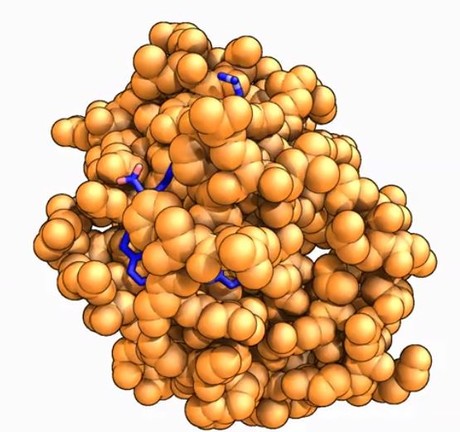
“It shows the three-dimensional structure of proteins, which do most of the work in cells, identifying opportunities to neutralise those involved in cancer and promoting those that may protect us from cancer.”
Australian Synchrotron Director Professor Andrew Peele added that the detector will more than double the facility’s capacity to collect data, leading to more targeted and effective treatments and, ultimately, improved patient outcomes.
“This new capability will take a beamline that was previously at full capacity — booked for use at all available hours of the day — and find it an extra gear, so it can deliver more research, and arm researchers with clear representations of protein structures,” said Professor Peele.
“We’re essentially shifting from dial-up internet to high-speed broadband, putting our foot on the accelerator of cancer research technology, providing faster protein analysis to turbocharge cancer research and facilitate significant discoveries.”
Water-based paints contain potentially hazardous chemicals
Some water-based paints contain compounds that are considered volatile organic compounds, along...
Portable device enables regular testing for kidney disease
Accessible, affordable urinalysis to assess kidney function could be a boon in remote areas as...
A major breakthrough in ultraviolet spectroscopy
Scientists successfully implemented high-resolution, linear-absorption dual-comb spectroscopy in...


