3D printing human tissues

Using a combination of ‘smart’ polymeric water-based gels and biodegradable plastics, a team of US-based scientists has 3D-printed muscle, bone and cartilage that has survived, matured and developed functional blood vessels when implanted in mice.
Developed over 10 years with funding from the Armed Forces Institute of Regenerative Medicine, the Integrated Tissue and Organ Printing System (ITOP) uses a custom-designed 3D printer built by a team of regenerative medicine scientists at Wake Forest Baptist Medical Center in Winston-Salem.
Previous attempts to 3D-print human tissues have failed due to a lack of blood vessels, but the ITOP tissues have been made with a lattice of microchannels that allow oxygen and nutrients to penetrate throughout the structures and eventually develop a system of blood vessels. The water-based ‘ink’ used in the 3D printing process has been optimised to promote cell growth and survive the printing process, while the plastic-like materials that form the strong but temporary outer structure are biodegradable. These factors help the implanted structures survive long enough to integrate with the body, a major challenge in tissue engineering.
Dr Anthony Atala, senior author of the study and director of the Wake Forest Institute for Regenerative Medicine, said: “Our results indicate that the bio-ink combination we used, combined with the microchannels, provides the right environment to keep the cells alive and to support cell and tissue growth.”
The ultimate goal of the project is to use data from CT and MRI scans to bioprint tailor-made muscle, bone and cartilage for individual patients to replace diseased or injured body parts.
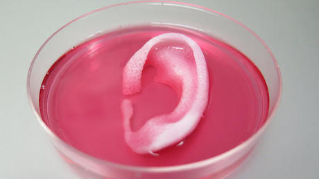
The latest proof-of-concept experiments utilising the ITOP system bioprinted human-sized ears, which were implanted under the skin of laboratory mice. Within two months, blood vessels and cartilage tissue had formed while maintaining the shape of the implanted ear.
Similar tests with bioprinted muscle tissue showed vascularisation and nerve formation within two weeks, while bioprinted jaw bone fragments formed vascularised bone tissue after five months.
Closer to home, scientists at the University of Wollongong’s Australian Institute for Innovative Materials have been 3D printing artificial human brains in order to study the mechanics of human-specific diseases such as schizophrenia and also developing a growth factor-rich bio-ink from seaweed extracts. Already proving useful in regrowing damaged cartilage, the next stage is to bioprint tissues that can mimic human organs.
Platform helps accelerate synthetic and metabolic workflows
Ultrahigh-throughput, droplet-based screening had long been on Biosyntia's radar for its...
Liquid biopsy to inform non-small cell lung cancer treatment
A novel liquid biopsy test may help determine which patients with non-small cell lung cancer that...
Small-scale bioreactor helps create cultivated meat
Magic Valley scientists have utilised an Applikon bioreactor in the process of creating...


