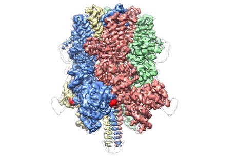
Atomic structure revealed for blood flow-regulating protein

US scientists have revealed for the first time the atomic-level structure of a promising drug target for conditions such as stroke and traumatic brain injury, with the results published in the journal Nature. Called TRPM4, this protein is found in tissues throughout the body, where it plays a major role in regulating blood flow via blood vessel constriction as well as setting the heart’s rhythm and moderating immune responses.
“Understanding the role TRPM4 plays in regulating circulation is vital, but for years research has been limited by a lack of insight about its molecular architecture,” said Wei Lü, an assistant professor at the Van Andel Institute (VAI) and lead author on the study.
TRPM4 is critically involved in regulating the blood supply to the brain, which comprises only about 2% of the body’s total weight yet receives 15–20% of its blood supply. Conditions that disrupt blood flow in the brain — such as stroke, traumatic brain injury, cerebral edema and hypertension — can have devastating consequences.
“Many safeguards exist in the brain’s circulatory system to protect against a sudden interruption in blood supply — one of which is TRPM4,” Lü said. “We hope that a better understanding of what this protein looks like will give scientists a molecular blueprint on which to base the design of more effective medications with fewer side effects.”
The structure of TRPM4 is markedly different from the other molecules in the TRP superfamily — a category of proteins that mediate responses to sensations and sensory stimuli such as pain, pressure, vision, temperature and taste. Broadly known as ion channels, proteins like TRP nestle within cells’ membranes, acting as gatekeepers for chemical signals passing into and out of the cell.
The Nature study represents the first atomic view of a member of the TRPM subfamily, revealing a crown-like structure with the four peaks composing a large N-terminal domain — a hallmark of TRPM proteins. This region, found at the start of the molecule, is a major site of interaction with the cellular environment and other molecules in the body. On the opposite end of TRPM4, commonly called the C-terminal domain, Lü’s team found an umbrella-like structure supported by a ‘pole’ and four helical ‘ribs’ — characteristics that have never before been observed.
“Our findings not only provide a detailed, atomic-level map of this critical protein, but also reveal completely unexpected facets of its makeup,” said Lü.
The findings were made possible by VARI’s David Van Andel Advanced Cryo-Electron Microscopy Suite, which allows scientists to view some of life’s smallest components in exquisite detail. VARI’s largest microscope, the Titan Krios, is one of fewer than 120 in the world and is so powerful that it can visualise molecules 1/10,000th of the width of a human hair.
A pre-emptive approach to treating leukaemia relapse
The monitoring of measurable residual disease (MRD), medication and low-dose chemotherapy is...
Long COVID abnormalities appear to resolve over time
Researchers at UNSW's Kirby Institute have shown that biomarkers in long COVID patients have...
RNA-targeted therapy shows promise for childhood dementia
Scientists have shown that a new RNA-targeted therapy can halt the progression of a specific type...


