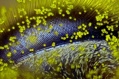
Beauty is in the eye of the bee

What’s the secret behind taking the winning image in a prestigious photomicrography competition? According to Queensland high school teacher Ralph Grimm, “It takes tons of patience, more than anything else.”
Since 1974, the Nikon Small World Photomicrography Competition has recognised the art, skill and scientific value of photography through the microscope. When evaluating the entries in the 2015 competition, the judges found themselves observing everything from chemical compounds to up-close-and-personal looks at biological specimens.
“Each year we are blown away by the incredible quality and quantity of microscopic images submitted from all over the world, from scientists, artists and photomicrographers of all levels and backgrounds,” said Eric Flem, communications manager, Nikon Instruments.
“Judges had their work cut out for them in narrowing down from such a rich pool of applicants, and we are so pleased with the results.”
Grimm was the lucky winner of this year’s top prize — $3000 to put towards the purchase of Nikon equipment — thanks to his stunning close-up of a honey bee eye covered in dandelion pollen grains. The image beat out more than 2000 entries from over 83 countries around the world, which was quite a coup for the self-taught photomicrographer.
“When I received the phone call that I won first place, I felt like I was dreaming,” Grimm admitted.
“I am a long-time participant [in the competition] and have entered my work, I think, since 1999.
“I have always admired the first-place winners of previous years, but never believed that I would be one of them.”

Grimm was especially aware of the fact that other entrants would have had “state-of-the art fluorescence and confocal technology available to them”. Yet the judges found that Grimm’s work demonstrated not only artistic quality but also exceptional scientific technique, with the image having taken over four hours to perfect.
“You may call it a labour of love,” Grimm said. “The specimen needs to be fresh, then it has to be secured and mounted on a rotatable base with a pivot so that its angle and position can be changed for composition. After that, the specimen is placed under the microscope.
“Because I’m not using a stereo microscope I have much less depth of field, and working distance is much shorter as well — which makes illumination a bit tricky if you don’t have an epi-illuminator. But the most tedious part of this photographic process is actually taking the focus stack, because if not done properly, it will lead to blank areas without any image information. After this is done, the final composite will have to be post processed in Photoshop or Lightroom.”
Even after this painstaking process, Grimm admitted that he originally didn’t want to submit the image of the honey bee eye at all. But not only had he invested a great deal of time and effort in his creation, it also highlighted an issue which is particularly close to the former beekeeper’s heart.
“I wanted to send a message out into the world,” said Grimm, “and it came to me that CCD, or colony collapse disorder, represents a serious problem with the honey bee — which is not only of economic concern, but ecological concern as well.
“Anyone can find out about CCD online, but the broader message is that if we don’t find a sustainable balance between commerce and the ecology, in the end nature will win. If the honey bee dies out, our lives will no longer be worth living. Food diversity will be severely reduced, and because 70% of crops need bee pollination, the agricultural industry will experience mega losses.
“A lot of factors seem to play together in the decline of honey bee populations — such as new types of pesticides, increasing parasite infestation and lack of genetic diversity — but I am particularly worried about the way humans rapidly change the environment. There is a lot of ignorance about this situation, especially here in Australia, with the building and construction madness shamelessly making room for city expansion and population growth. It’s my little way of expressing my fear. The bee, to me, is like a sweet prophet with a sour message.”
Of course, Grimm was not the only photomicrographer highlighted in the competition. Coming in second place was the joint effort of Kristen Earle, Gabriel Billings, KC Huang and Justin Sonnenburg from the Stanford University School of Medicine, USA. Their image displays the colon of a mouse that was born germ-free. The mouse was colonised with a human microbiota and used DNA probes to label certain taxa — in this case, members of the Bacteroidetes and Firmicutes phyla.

In third place was Dr Igor Siwanowicz from the Howard Hughes Medical Institute (HHMI), USA. His image shows the entrance to the trap (or bladder) of the humped bladderwort (Urticulatia gibba), a carnivorous freshwater plant. Several elements of the bladder’s construction are visible in the image, giving some insight into the workings of this elaborate, 1.5 mm-long suction trap.

All the winners of this year’s competition — including the Top 20, 12 Honourable Mentions and 56 Images of Distinction — can be found at www.nikonsmallworld.com/galleries/photo. Grimm hopes the images will be of interest to the general public as well as scientists, saying, “The beauty of nature captured by a photographer should evoke a sense of awe in viewers without a scientific background.”
Nikon will also unveil the winners of its sister competition, Nikon Small World in Motion, later this year. Showcasing the best of science and art under the microscope in the form of video, the 2015 competition winners will be revealed on 2 December on www.nikonsmallworld.com/galleries/swim.
“Nikon is doing a wonderful job by giving the work of photomicrographers around the world a chance to be noticed by a larger audience,” said Grimm.
Smart nanoprobe lights up prostate cancer cells
Researchers have developed a smart nanoprobe designed to infiltrate prostate tumours and send...
DESI's 3D map more precisely measures the expanding universe
The Dark Energy Spectroscopic Instrument (DESI) has created the largest 3D map of our cosmos ever...
Toxic metal particles found in cannabis vapes
Nano-sized toxic metal particles may be present in cannabis vaping liquids even before the vaping...


