Improving X-ray spectrometers

Swiss researchers have developed a new design for X-ray spectrometers that lowers overall production costs and increases the efficiency of X-ray flux. This may lead to faster acquisition times for sample imaging and increased efficiency, which is important for biological samples that may be damaged by continued X-ray exposure.
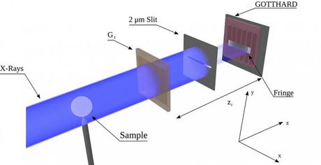
X-ray interferometry works by firing X-rays at a downstream detector. Along the way, the beams pass through a phase grating, which divides the beam into different diffraction orders based on their wavelength. The difference between these diffraction orders introduces an interference fringe — a source of interference which needs to be in the micrometre range in order to achieve high sensitivity for the detector. Such fringes are challenging to record directly over a large field of view, necessitating the use of highly sensitive detectors.
In response to this, a method known as Talbot-Lau interferometry was developed and adopted. The method utilises an absorption grating, G2, placed right before the detector, and senses the distortions via phase stepping. Here, the absorption grating is scanned step by step for one or more periods of the interference fringe, each time recording an image which results in an intensity curve at each pixel. This allows the interference fringe to be sensed indirectly, while obtaining absorption, differential phase and small-angle scattering signals for each pixel.
However, this ultimately causes the system to be less efficient for each dose of X-rays due to photon absorption by G2. The required area and aspect ratio of the gratings, which are millimetre-sized, further complicate matters by driving up overall production costs.
To remedy this, researchers at Zurich’s Institute for Biomedical Engineering and the Paul Scherrer Institute’s (PSI) Swiss Light Source (SLS) developed an interferometer that does not use the G2 grating and instead directly exploits the fringe interference for higher resolution. Their research has been published in the journal Applied Physics Letters.
The researchers’ set-up consisted of an X-ray source, a single phase grating and a microstrip detector developed by the SLS detector group — a simplified version of the traditional Talbot-Lau interferometer. The detector uses a direct conversion sensor in which X-ray photons are absorbed and the charge generated from one absorption event is collected by more than one channel for small channel sizes — charge sharing.
“The key point to resolving the fringe is to acquire single-photon events and then interpolate their positions using the charge sharing effect, which is usually considered as a negative effect in photon counting detectors,” said Matias Kagias, a co-author on the paper. By interpolating the position of many photons, a high-resolution image can be acquired.
When the researchers implemented the appropriate algorithm to analyse this recorded fringe, they found that fringes of a few micrometres could be acquired successfully while still retrieving the differential phase signal. According to Kagias, this increases the interferometer’s flux efficiency by a factor of two compared to a standard Talbot-Lau interferometer, which may lead to faster acquisition times and a dose reduction.
A non-destructive way to locate microplastics in body tissue
Currently available analytical methods either destroy tissue in the body or do not allow...
Rapid imaging method shows how medicine moves beneath the skin
Researchers have developed a rapid imaging technique that allows them to visualise, within...
Fluorescent molecules glow in water, enhancing cell imaging
Researchers have developed a new family of fluorescent molecules that glow in a surprising way,...


