
Asbestos testing under the microscope

Asbestos is a naturally occurring silicate (silicon- and oxygen-containing) mineral that has a fibrous structure. These fibres are composed of microscopic fibres that can become airborne when the asbestos is disturbed, making asbestos easily inhaled. As testing for its presence can be problematic, Professor Akio Kuroda from Hiroshima University has been working on a novel solution.
Formerly enjoying widespread use for its insulation and fireproofing qualities, asbestos has been banned in many countries since the discovery that asbestos fibre inhalation can cause lung cancer, asbestosis and other lethal lung conditions. Unfortunately, Kuroda noted, large quantities of asbestos-containing materials remain in old buildings, and people are at risk of exposure to asbestos during demolition.
“In addition, natural minerals such as talc, a raw material used in the manufacture of cosmetics, pharmaceuticals and baby powder, may also contain asbestos,” Kuroda said. “It has recently been highlighted that asbestos-contaminated talc may cause cancer.”
Aware of the importance of fast and accurate asbestos testing, Kuroda has been developing testing techniques to accurately pinpoint the presence of asbestos. His approach to establishing better testing for asbestos has been twofold, with the development of a fluorescent microscopy (FM) method that offers increased sensitivity and convenience, as well as the creation of an asbestos-specific protein probe combined with a fluorescent marker that allows users to easily visualise asbestos fibres under a fluorescent microscope.
A need for improved testing
Testing for asbestos is currently often accomplished using polarised light microscopy (PLM). While this method is convenient and cheap to run, the sensitivity is insufficient for the detection of fine fibres. The US FDA recommends transmission electron microscopy (TEM) due to its higher sensitivity; however, it is labour-intensive as it requires samples to be sent to a laboratory to be processed, and therefore testing becomes more costly. Phase contrast microscopy (PCM) can be carried out in situ, enabling faster results, but is not able to distinguish between asbestos and other types of fibres.
Kuroda’s approach has been to target FM as a highly specific way to identify asbestos, with a 2012 study from the US National Institutes of Health finding that FM was able to detect fibres that were almost 10 times smaller than those found by PCM testing. The most difficult challenge he faced, having developed a specific probe for asbestos, has been to quantify what percentage of fluorescent fibres made visible in the test samples are asbestos.
“To verify the identity of the fibres that were fluorescently tagged, we analysed them under an electron microscope to identify their elemental composition,” Kuroda said. “However, it was very difficult to find the individual fibres under both microscopes in order to verify them.”
In a sample that may contain thousands or even hundreds of thousands of fibres, this task was like finding the proverbial needle in a haystack. Fortunately, Kuroda was able to turn to recent advances in imaging technology to solve this issue.
“We were able to use a newly developed correlative microscopy system where the sample stage is shared among the two microscopes,” he said, which means that the location information of fibres therefore remained. Kuroda said the results were astounding — over 95% of fluorescent fibres observed in the practical samples were really asbestos.
By creating a probe that is highly specific for asbestos, by modifying the asbestos-binding protein with fluorescent dye, it is now possible to visualise this lethal substance in samples of construction materials, cosmetics and even baby products. Before this finding, Kuroda said, no one knew that asbestos could be fluorescently visualised.

With results published in the Annals of Work Exposures and Health (previously The Annals of Occupational Hygiene), Kuroda’s methods were approved in the Asbestos Monitoring Manual, a guide published by Japan’s Ministry of the Environment. The probes he developed in this study have been made commercially available and are currently used by several companies for fast and accurate monitoring of asbestos.
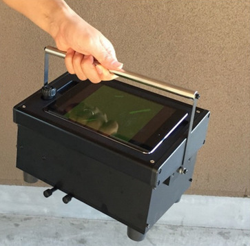
In addition to developing the probe and validating his results, Kuroda has also worked with a commercial manufacturer of microscopes to create a fluorescent microscope that was suitable for use in situ. Existing fluorescent microscopes were unwieldy devices that needed to be used in a laboratory setting and were quite unsuitable for transport around dusty demolition sites, but Kuroda and his industry partners were able to design a robust and portable microscope that could be taken onsite, thus making testing faster and more accurate.
By tackling multiple problems that had beset the arena of asbestos testing and control, Kuroda has brought faster and more accurate testing to contaminated sites worldwide. This means greater safety not just for construction workers labouring on potentially hazardous sites, but also for all citizens living and working in polluted homes and buildings.
Please follow us and share on Twitter and Facebook. You can also subscribe for FREE to our weekly newsletters and bimonthly magazine.
Prototype promises to build light analysis directly into imaging hardware
Thanks to a new chip, researchers believe they may be able to help cameras capture details that...
Ultrasound improves protein recovery from cauliflower waste
Abundant but often discarded during processing, cauliflower leaves contain protein and dietary...
Can yawning advance the study of neurodegenerative diseases?
Australian neuroscientists using MRI scans to monitor flow of cerebrospinal fluid during yawning...


