3D 'video' reveals inner activity of cells
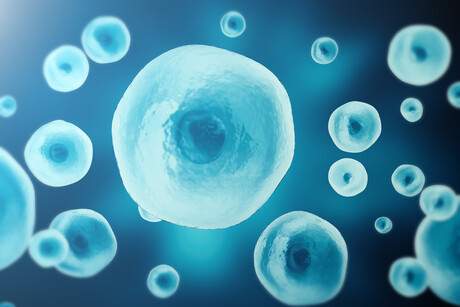
Researchers at The University of Tokyo have developed a high-speed microscopy technique which captures images so quickly they can be used to make ‘videos’ of the dynamic processes which occur inside living cells. Their work has been described in the journal Nature Communications.
There are many ways to look inside our cells and see how they work, adapt to change and respond to treatments. One technique is label-free mid-infrared photothermal microscopy. This uses mid-infrared light to vibrate the chemical bonds within molecules, each of which has a distinctive frequency. The different frequency signatures can be used to create a map of the internal composition of a cell, which can then be seen by illuminating it with visible light.
An advantage of this technique is that, unlike with some other methods, no dyes are used which can be toxic to live samples, and by combining shots from multiple angles, a 3D picture can be created showing active biological processes in motion. However, capturing images at subsecond timescales has proven challenging.
“Previously, three-dimensional mid-infrared photothermal tomography was limited to less than 0.05 images per second, mainly because signal averaging and sequential measurements from different illumination angles required considerable time,” said PhD student Masato Fukushima, first author on the new study. “However, we have developed a new technique which achieves 19.2 images per second, enabling us to acquire a complete three-dimensional image every 52 milliseconds (thousandths of a second) — approximately 400 times faster than previous approaches.”
The team overcame the speed barrier by using a spatial light modulator (a device that precisely shapes the properties of light, such as its wavelike phases of crests and troughs, to steer it in different directions) to switch the illumination angle very quickly, and by maximising the signal-to-noise ratio for each shot to make it as clear as possible. The resulting system is called video-rate mid-infrared photothermal optical diffraction tomography (MIP-ODT).
Using MIP-ODT, the team could view lipid droplets in a cell (which are responsible for regulating energy and storing lipids) and track how they moved in three-dimensional space, revealing transport behaviours which could not easily be seen through 2D imaging alone.
“Our research transforms this technique from a method primarily used to observe relatively static samples into a tool capable of quantitatively investigating fast biological processes in three dimensions,” Fukushima said.
The researchers believe this new technique will enable them to address fundamental biological questions that cannot be explored with conventional methods. One use could be the study of biomolecular condensates — membraneless structures in cells which contain proteins and nucleic acids. Abnormal behaviour of these condensates has been linked to neurodegenerative disorders, so investigating how they grow and change may help researchers develop new treatments.
“A key strength of label-free chemical imaging is its ability to provide quantitative information about the chemical composition of living cells,” said senior author Professor Takuro Ideguchi. “By visualising cellular dynamics in three dimensions and over time, we aim to advance toward a quantitative, measurement-driven understanding of life.”
Micromirrors improve image quality for optical microscopes
A specially designed micromechanical mirror array corrects optical distortions that occur when...
Method holds promise to make bioimaging X-ray machines smaller and more flexible
Researchers from Nanyang Technological University, Singapore have found a new way to produce...
A non-destructive way to locate microplastics in body tissue
Currently available analytical methods either destroy tissue in the body or do not allow...