Micromirrors improve image quality for optical microscopes
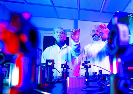
Researchers at the Fraunhofer Institute for Photonic Microsystems IPMS have developed a new technology that can significantly improve the image quality of optical microscopes. Their specially designed micromechanical mirror array corrects optical distortions that occur when viewing biological tissue, making fine structures appear sharper — an important prerequisite for biomedical research and future applications in medical diagnostics.
One of the biggest challenges in microscopy is light scattering, as biological tissue distorts light as it passes through the sample. This results in blurry images and a limited depth of field, making high-resolution imaging particularly difficult with living samples.
To compensate for these distortions, a technique known as adaptive optics is used. It specifically modifies the wavefront shape of the light, thereby correcting image aberrations. However, this has typically required additional sensors or complex optical systems.
“Until now, microscopes had to first measure the image aberrations and then correct them,” said Dr Maxim Darvin, Project Manager at Fraunhofer IPMS. “Our new mirror array performs both tasks simultaneously, which saves time and simplifies the entire set-up.”
At the heart of the development is a chip containing approximately 64,000 movable micromirrors. Each individual mirror measures just 16 µm, meaning that four such mirrors can fit side by side on the thickness of a human hair. The micromirrors can be adjusted to focus light through scattering materials with high contrast, thereby enabling the analysis of structures in deeper tissue layers at higher resolution.
Each mirror can be adjusted with extreme precision to 256 different positions without tilting and changes its alignment up to 1000 times per second, allowing the system to compensate for optical disturbances with virtually no delay. The system is suitable for wavelengths ranging from deep ultraviolet to the near-infrared and is independent of the light’s polarisation, making it flexible and suitable for a variety of microscopy techniques.
“We have already demonstrated the performance of our development on various biological samples, including corneal cells and cellulose,” Darvin said. “In all cases, our adaptive optics delivered visibly sharper and higher-contrast images. We have also succeeded in precisely focusing the laser light through highly scattering materials. This allows structures in deeper tissue layers to be examined with higher image quality.”
Due to the algorithm, the system currently still takes a few minutes to fully identify and correct image distortions; the goal of further development is to perform these processes in near real time in the future. In the long term, this technology could help make high-resolution microscopy faster, simpler and more powerful, both in basic biological research and in future diagnostic applications in medicine.
Method holds promise to make bioimaging X-ray machines smaller and more flexible
Researchers from Nanyang Technological University, Singapore have found a new way to produce...
A non-destructive way to locate microplastics in body tissue
Currently available analytical methods either destroy tissue in the body or do not allow...
Rapid imaging method shows how medicine moves beneath the skin
Researchers have developed a rapid imaging technique that allows them to visualise, within...