Dry and resoak: the new way to speed up bioassays

Scientists from the University of Washington (UW) have proposed a way to speed up the time it takes to conduct biological assays. Their solution, reminiscent of the science behind washing machines, could reduce wait times from hours to minutes.
Many biological assays use molecules such as antibodies to detect specific types of cellular proteins or pieces of DNA. These ‘detector molecules’ only bind to specific targets, such as a certain class of cellular proteins, and include additional components such as nanoparticles or dye molecules to emit light if they successfully bind. These assays have revealed where different proteins are found in cells and helped diagnose diseases — but such tests can take hours or days to complete.
The detector molecules, suspended in a fluid, float around while their targets — whether cellular proteins or pieces of DNA — are adhered to the hard, flat surface of a small plate or Petri dish. While bulky detector molecules close to the surface can easily find and bind to their targets, molecules further up in the fluid column move slowly due to their size. It can take hours for enough detector molecules to diffuse down and bind to their targets to produce a visible colour change. This is called diffusion limitation.
UW Associate Professor Xiaohu Gao and his team decided to tackle the problem of diffusion limitation after they developed a new staining assay whose long reaction times made their protocol impractical. Instead of waiting for detector molecules to drift down to the surface of the plate, they allowed detector molecules close to the surface to bind. They then drained the solution from the plate, mixed it, put it back on the plate and repeated this cycle dozens of times — what they call cyclic solution draining and replenishing.
“In a washing machine, you squeeze water out and put it back in,” said Associate Professor Gao. “Dry and resoak. Dry and resoak. This is exactly the same mechanism: drain the fluid completely and then put it back on the plate. That’s much more efficient than simply stirring it around.”

To drain fluid from the plate, the team covered the plate with a seal and inverted it. To resoak, they flipped the plate upright again. The flipping action helped mix the detector molecules in the fluid, which sped up the total reaction time. And while sealing and flipping might be impractical for some tests, Associate Professor Gao noted that “you could use air bubbles or centrifugation to drain as well”.
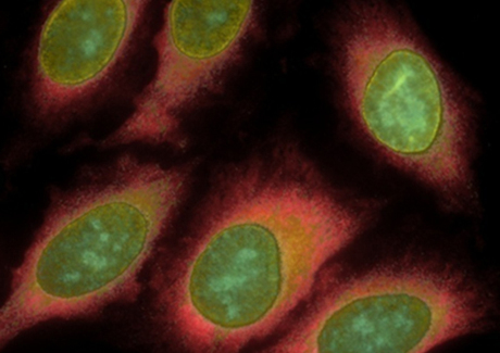
The team tested cyclic solution draining and replenishing with two antibody staining techniques: ELISA and immunofluorescence microscopy. Reaction times for both were cut substantially; in one case, a one-hour incubation time was cut to just seven minutes. The results of the study have since been published in the journal Small.
“When we prepare tea, we don’t let it sit there or shake the cup,” said Associate Professor Gao. “We repeatedly lift, drain the tea bag, then lower it into the hot water. That’s what we’ve done here.
“This was a common problem that no-one before had made a link to. But here we have, and it’s so simple.”
Blood test could be used to diagnose Parkinson's earlier
Researchers have developed a new method that requires only a blood draw, offering a non-invasive...
Cord blood test could predict a baby's risk of type 2 diabetes
By analysing the DNA in cord blood from babies born to mothers with gestational diabetes,...
DNA analysis device built with a basic 3D printer
The Do-It-Yourself Nucleic Acid Fluorometer, or DIYNAFLUOR, is a portable device that measures...


