
LiveCyte: Every cell tells a story

The Challenges of Live Cell Imaging
The biggest challenge for many live cell researchers is to characterise individual cells without impacting their behaviour through the act of monitoring them. Whilst the use of fluorescent labels allows both cells and cellular functions to be visualised, the levels of illumination needed to excite the fluorophores can alter innate properties of cells, with the associated cytotoxicity limiting the scope, duration and integrity of any experiment. Hence, the emergence of label-free techniques, which exploit the inherent contrast of cellular components to create images, that allows cells to be tracked and monitored over longer periods of time. However, for many such techniques, constraints caused by low levels of contrast preclude the automated segmentation of individual cells and by association the capability to differentiate them based on subtle differences in their morphology and motion.
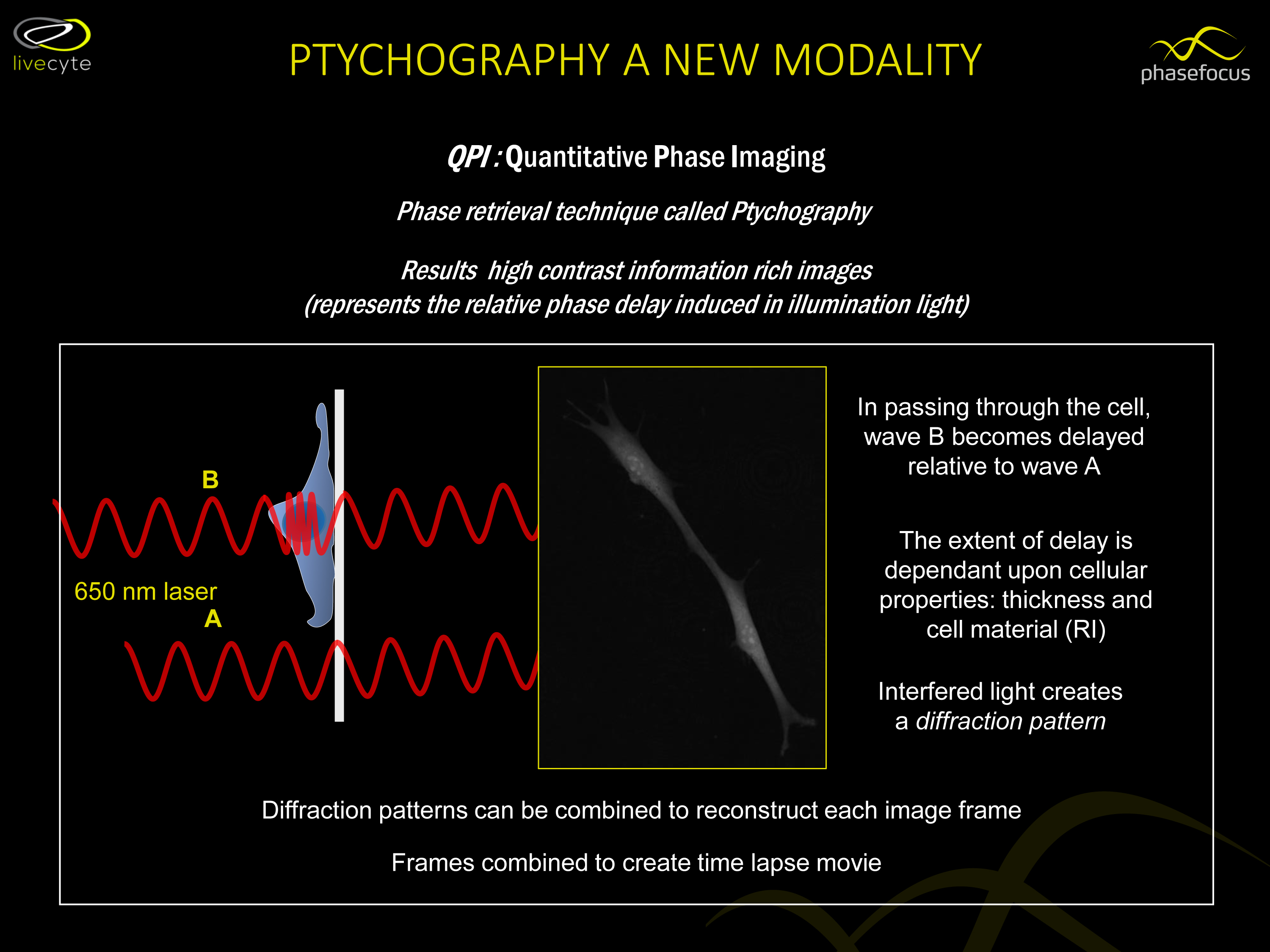
The LiveCyte™ imaging & cell analysis system from Phasefocus addresses these limitations by employing Ptychographic Quantitative phase imaging (QPI). This technology leverages phase shift information to generate high contrast cell images under low levels of light intensity, allowing individual cells to be identified and tracked for prolonged periods without the need for perturbing labels. This ability to image under a more natural environment with reduced risk of phototoxicity not only supports the use of sensitive cell types such as primary and stem cells, but also enables viable cells to be recovered for subsequent experimentation or downstream analysis, giving it broad spectrum appeal and for clinical applications in particular.
QPI: Making every cell count
The ability to segment and follow individual cells is paramount for accurate quantification of cell behaviour and differentiates LiveCyte from other commercially available live cell imaging systems.
Ptychography is a computational technique, and unlike optical methods where intensity variations make it difficult to extract quantitative data, the full extent of phase shift can be calculated and subsequently translated into high contrast, artefact free images. Furthermore, the technology employed in LiveCyte delivers a continuous field of view with no loss of resolution permitting even highly motile cells to be tracked during time-lapse imaging, ensuring no cells are “lost”.
It is the fidelity and quantitative nature of the images which enables the direct measurement of cell parameters, allowing specific morphological and dynamic characteristics of the cells to be measured. As a result, researchers not only gain a true record of cell count for the population as a whole but have the added capability to define and quantify distinct sub-populations within complex heterogeneous cultures, achieving a more realistic narrative of cell behaviour.
Automated, multi-parametric analysis
Superior time-lapse imaging is critical in its own right, but in order to ascertain the precise impact of environmental conditions, it is the data analysis that is the crux of every experiment. LiveCyte’s Cell Analysis Toolbox™ (CAT) contains automated tracking software that monitors changes in individual cells, through multiple cell divisions eliminating the need for manual tracking, achieving a seamless integration of image acquisition and analysis.
Each experiment automatically yields a plethora of metrics, providing information on phenotypic parameters such as cell thickness, volume, dry mass in addition to kinetic behaviour characterised by factors including cell speed, displacement and meandering index.
With the capability to compare response to different treatment or environmental conditions at both population and single cell level within a single experiment, laboratory workflows can be effectively streamlined, making the best use of limited resources.
In the current economic climate, as researchers face ongoing cost pressures and demands for productivity improvements, LiveCyte represents a rapid and cost-effective means of gaining deeper insights into biological processes, associated with a wide range of disease conditions with positive implications for drug discovery and development of personalised medicine.
ATA Scientific is pleased to be the local distributor for the LiveCyte Cell Imaging and Analysis system developed by UK-based company Phasefocus™.
Reference: Kasprowicz, R., Suman, R., O’Toole, P. Characterising live cell behaviour: Traditional label-free and quantitative phase imaging approaches. J. Biochem. & Cell Bio. 84 (2017) 89-95.
ATA Scientific Pty Ltd
+61 2 9541 3500
enquiries@atascientific.com.au
www.atascientific.com.au
For more information: https://www.atascientific.com.au/live-cell-imaging-analysis-using-quantitative-phase-imaging-qpi/.
For a larger version of the image, click here.
Celebrating 25 years in optical imaging expertise with over 30,000 IVIS publications!
It's been 25 years since the very first IVIS preclinical optical imaging system was launched,...
Efficient and Reliable Gas Control in Material Analysis with Burkert
Burkert's expertise in fluidics facilitates the development of tailored gas control solutions...
Delving deeper into CAR T cell evaluation
Discover a 3D imaging workflow to study the infiltration potential of CAR T cells into solid tumors.


