
See what you can accomplish with an Azure Biosystems Sapphire Biomolecular Imager

Whatever type of imaging your lab does — whether it’s the ubiquitous western blot, southern blots of 2D DNA gels, visualizing gross morphology of tissues or small model animals, or something more unique — the Sapphire™ Biomolecular Imager from Azure Biosystems will deliver outstanding, quantitative detection with NIR and RGB fluorescence, chemiluminescence, and phosphor imaging.
The highly capable, multi-functional Sapphire Biomolecular Imager is here to simplify your search and save you time. Instead of looking for multiple instruments to document and quantify visual data, such as bulky camera and lightbox setups, film cassettes and developers, and separate gel and western blot documentation systems, all these capabilities and more can be done with a single bench-top instrument.
With the ability to acquire and analyse RGB and NIR fluorescence, chemiluminescence at the same sensitivity as film, and data from phosphor image screens, the Sapphire gives you the ability to do so much with a single instrument.
Applications include:
- Quantitative western blotting
- NIR western blotting
- Chemiluminescence
- Protein gel documentation
- DNA gel documentation
- In-gel imaging/in-gel western
- Protein array imaging
- EMSA
- 2D-DIGE
- Reporter gene assays
- Tissue imaging/Tissue section imaging
- Small model organism imaging
- Phosphor imaging
- In-cell/on-cell ELISA
- 2D DNA gels
- MORE

WESTERN BLOTS:
What makes the Sapphire so great for sensitive detection and quantitation of chemiluminescent and fluorescent western blots?
Four solid-state lasers deliver strong excitation and the unique, three-detector design maximizes performance by ensuring that the right sensor is used for the type of imaging being done. A sensitive photomultiplier tube (PMT) optimizes blue light detection and phosphor imaging. A high quantum-efficiency avalanche photodiode (APD) enables near infrared (NIR), infrared (IR), red, and green light imaging. While a CCD sensor provides chemiluminescent imaging with the same sensitivity as film. The Sapphire Biomolecular Imager comes with easy-to-use Sapphire Capture and AzureSpot analysis software.
Fluorescent western blotting tip:
- Improve sensitivity by drying your western blot before imaging.
While scanning a wet membrane does produce detectable signal, drying the membrane results in increased signal intensities, lower background and better signal to noise ratios. Water can attenuate fluorescence and even slight differences in the dryness of different regions of a blot can lead to variable quantitation. Drying your blot prior to imaging can greatly improve sensitivity and the ability to generate reliable quantitative data.
See example below:

GEL IMAGING:
The same three detector technology that makes the Sapphire so great for imaging western blots is also flexible enough to image a wide range of gels, whether they are ethidium bromide (EtBr)-stained DNA agarose gels, coomassie-stained protein gels, or even 32P-labeled DNA acrylamide gels.
- Measure protein-DNA binding using EMSA. Image delicate gels while still in glass plates
- View and quantify Sypro Ruby-stained 2D protein gels. Analyze proteomics studies with ease
- View and quantify 35S-labeled proteins in 2D gels. Perform proteomics analysis on metabolically-labelled samples
- Image coomassie- and silver-stained protein gels
- Get accurate DNA quantitation from EtBr-stained agarose gels
- Image Midori Green-stained DNA agarose gels
- Directly detect DNA for Sanger sequencing and foot printing
TISSUE & SMALL ANIMAL MODEL IMAGING:
With the capability to image down to 10 μm resolution and a 25 cm x 25 cm scanning bed, the Sapphire Biomolecular Imager can go from scanning blots to scanning tissues and small animal models like mice, rats, small plants, and zebrafish. Quickly capture and quantify gross anatomy, morphology, protein localization and much more.
Track protein movement through tissue in small animals. Use the Sapphire Biomolecular Imager to image whole small animal models using fluorescence, chemiluminescence, and phosphorimaging detection.
- Get information on tissue structure. Using CLARITY for whole brain imaging
- Measure protein localization in tissue. Studying the permeability of embryo/placenta barrier
- Visualize anatomical structures in rats, mice
- Image Xenopus oocytes, embryos and track protein localization with fluorescence
- Track viral infection and quantify viral load. The Sapphire™ can be used to track localization of more than just protein.
Below, FITC-labeled virus is used to infect a zebrafish, which is then placed directly onto the Sapphire for imaging.

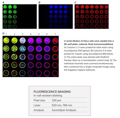
96-WELL IMAGING
- Image cells in multi-well plates. Measuring cell viability using crystal violet
- Improve efficiency with in-cell western blotting. Accurately quantify intracellular proteins with the repeatability, speed, and throughput of an ELISA
SOUTHERN BLOTS:
With the ability to image radiolabelled, fluorescently-labelled, and even chemiluminescently-labelled molecules, the Sapphire places an array of southern blot detection technologies at your fingertips.
- Measuring plasmid abundance with both phosphorimaging and chemiluminescence.
- Sensitive, quantitative DNA detection with a 32P-labeled probe.
- Determining DNA structure with 2D agarose gel electrophoresis.
- Measuring light chain-heavy chain DNA ratios for recombinant antibody expression.
GENERAL IMAGE CAPTURE:

Contact: Scitech Pty. Ltd
PO Box 1625
Preston South VIC 3072
Australia
Office: (03) 9480 4999
E-mail: sales@scitech.com.au
Website: www.scitech.com.au
Celebrating 25 years in optical imaging expertise with over 30,000 IVIS publications!
It's been 25 years since the very first IVIS preclinical optical imaging system was launched,...
Efficient and Reliable Gas Control in Material Analysis with Burkert
Burkert's expertise in fluidics facilitates the development of tailored gas control solutions...
Delving deeper into CAR T cell evaluation
Discover a 3D imaging workflow to study the infiltration potential of CAR T cells into solid tumors.


