Better blood vessel imaging detects cancer early

Researchers at CSIRO’s Data61 are developing a software tool that could significantly improve the detection of the development of new blood vessels, which is known to precede the growth of cancers. Earlier detection of blood vessel growth may lead to a faster diagnosis of malignant tumour growth, which is a key factor in successful treatment and patient survival.
As explained by Cancer Council Australia CEO Professor Sanchia Aranda, the capacity of cancers to form new blood vessels (angiogenesis) is a critical feature enabling cancer cells to spread to other parts of the body. Anti-angiogenesis treatment aims at prevent cancers from growing blood vessels.
The ability to continuously monitor subtle proliferations in blood vessels over time is essential, given that patients may react to anti-angiogenesis treatment differently. The accurate quantification of vasculature changes, particularly the number of terminal vessel branches, can play a critical role in accurate assessment and treatment.
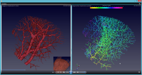
Images of blood vessel structure taken from high-resolution imaging have until now only produced a skeletonised view of blood vessel structure, which provided limited detail and accuracy. The researchers at Data61 are looking to change all that, teaming up with the Shanghai Institute of Applied Physics, Chinese Academy of Sciences to produce images of the brains and livers of mice at various stages of cancer growth.
The researchers analysed 26 high-resolution 3D micro-CT images from 26 mice, produced by the Shanghai Synchrotron Radiation Facility (SSRF). Using the images, the team developed a robust algorithm to generate an accurate representation of the vasculature, preserving the length and shape information of the blood vessel and its branches.
The new software allows researchers to measure subtle changes in the proliferation of blood vessels, including the number and length of the blood vessel branches, and produces significantly clearer skeletons of the vasculature than previously possible. According to Professor Aranda, the project “seeks to bring to life the tumour micro-environment through 3D synchrotron images of the vessels and will help to advance our understanding of this critical cancer progression process”.
“The hope is that through improved understanding, new opportunities to disrupt angiogenesis will be identified and open pathways to new treatments,” she said.
While the development of this new technique is a significant step forward, the Shanghai Synchrotron Beamline used to produce the images generates radiation levels unsafe for human imaging. In order to progress clinical trials in humans, the researchers are partnering with a hardware manufacturer that can produce high-resolution 3D images with safe levels of radiation for humans.
Dr Dadong Wang, lead researcher on this project from Data61’s Quantitative Imaging team, said that the applications of the software could be extended beyond 3D angiogenesis analysis to a wide range of other applications, such as analysis of 3D neurite outgrowth for drug development.
“We are very hopeful,” he said, “and currently looking for collaborators and partners to take the technology to the next stage.”
Beckman Coulter Life Sciences announces Automata partnership
Beckman Coulter Life Sciences has announced a strategic partnership with Automata — to...
AI spots hidden objects in chest scans
A new artificial intelligence (AI) tool can spot hard-to-see objects lodged in patients'...
Drying biofluid droplets used to diagnose disease
A newly developed workflow relies on the drying process of biofluid droplets to distinguish...


