
From cancer tissue to dying stars: Extreme Imaging 2014
Friday, 28 March, 2014
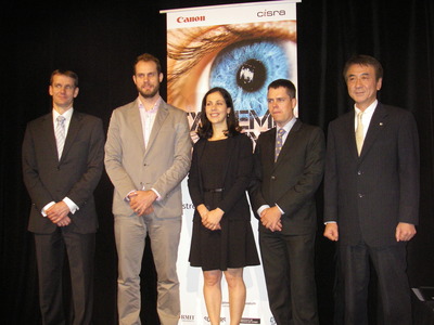
The winners of the 2014 Extreme Imaging Competition were last night announced at the Powerhouse Museum. Now in its third year, the annual event is held by Canon Australia’s local research and development arm, Canon Information Systems Research Australia (CiSRA).
As explained by CiSRA R&D Manager Roger Butler, the competition was originally set up to keep track of Australian imaging research, in particular that coming out of our universities. He added that the research facility - claimed to be Australia’s largest outside of Japan - uses the entries to find possible recruits to its world-class group of researchers and engineers.
“CiSRA remains at the forefront of digital imaging by developing and maintaining expertise in the computational and algorithmic aspects of image enhancement, image transformation and image analysis,” Butler said.
Dr Geoff Woolfe, Senior General Manager, Image and Video Research Centre CiSRA, said the event now strives to encourage students to go above and beyond the realm of conventional imaging. It is open to all Australian university students who are developing a new technique or equipment which produces images as part of their research project.
This year’s winner was Loretta Scolaro, of the University of Western Australia, who was supervised by Professor David D Sampson. Scolaro’s project set out to find a quick, minimally invasive method of imaging breast cancer in the body, addressing the fact that when breast cancer tissue is cut out during surgery, the surgeons don’t exactly know the boundaries of the tumour.
“If it’s found that the analysed, excised tissue contains tumourised boundaries, it’s likely that cancer cells were left behind in the body,” said Scolaro. This means patients will have to come in for a second round of surgery, she said, and currently occurs in 26% of cases.
“How can we image cancer at a microscope level which allows us to see its boundaries while still being in the body?” Scolaro asked. She and her team set out to use optical coherence tomography (OCT) - a relatively new technique which is analogous to ultrasound, easy and fairly inexpensive, but can only see 1 to 2 mm in depth. The researchers thus looked to developing an optic probe that could be inserted inside a needle, which could be delivered to the site of the cancer inside the body.
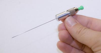
Scolaro said the needle, probe and accompanying lenses and mirror had to be “small, to be minimally invasive; robust, to be used during surgery; and easy to manufacture”. The final needle was made to be just over half a millimetre in width, and the researchers are now looking at utilising it in pilot clinical studies.
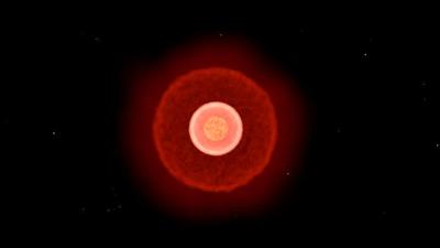
The runner-up was Paul Stewart, of the University of Sydney, supervised by Dr Peter Tuthill. Stewart proclaimed, “I’m going to show you how you can use space robots to turn the rings of Saturn into a giant telescope.”
He explained that the NASA spacecraft Cassini has been orbiting Saturn for around 10 years, and the craft will “watch a star as it passes behind the rings with a spectrograph”. As a star passes behind the planet’s rings, its light source is diffracted around the rings’ edges, because they are so sharp. “Each edge within the rings will actually give you a slice through the star in a different direction normal to the edge at that point,” explained Stewart.
“Once you have the brightness profiles from each direction, you can actually project them over each other in the same way that is done in medical tomography, and then apply regression algorithms which will take out a lot of the artefacts and essentially reduce the complexity of the image to get you an image that’s closest to what the true image of the star would be.”
Winner of the undergraduate prize was Charles Baker from The University of Queensland, supervised by Dr Nicholas Dowson, Dr Paul Thomas and Professor Steven Rose. Baker has developed a kinetic model to image liver lesions that takes into account the structural and functional features of the liver.
Baker explained that during a PET (positron emission tomography) scan, the regions of the body are imaged as a collection of 3D cubes. Over a period of around an hour, the patient has to lie motionless while the PET scanner fires up a number of images, eventually generating a static PET image.
“Having a patient in a PET scanner for 60 to 75 minutes is not easy to do,” Baker noted. The project thus had several aims, he said: to image smaller lesions; to determine the bioactive areas of larger lesions; to reduce nuclear medicine reporting time; and to reduce the total scan time.
Butler said this was his second year judging the competition and that the entries get stronger every year. Among those entrants which didn’t reach the finals, he listed technology for detecting smoke in video footage; a terahertz lens capable of propagating sub-wavelengths over long distances; and a high-speed camera, attached to an X-ray synchrotron, which captured images of lung tissue.
“I’d like to thank and congratulate the students and their supervisors who entered the competition, not just the finalists but all of them; they were all really good quality, and separating them was quite difficult,” Butler said. “We were pleased to receive a very interesting and diverse range of quality applications; the judging really was a delight as well as a challenge.”
Scolaro said it was “fantastic” just to be a part of the awards night.
“It’s inspired, I think, a lot of the research, and just to see the quality of research in other fields has been a privilege,” she said. “It’s a lot to take in. So many quality entries.”
Woolfe concluded, “It is our hope that this competition continues to encourage and support some of the best Australian research in imaging and leads to even greater participation in imaging-related research in the country.
“It’s a privilege for Canon to be able to support the next generation of leading Australian scientists.”
How librarians can help maintain image integrity
By sharing best practice and providing useful resources, librarians can support researchers in...
Winners announced for 2025 Australian Museum Eureka Prizes
Now in their 35th year, the Australian Museum Eureka Prizes continue to highlight some of the...
"Damning" review of Forensic Science Queensland released
The review of the troubled forensic service provider reveals unreliable results,...


