
A mini microscope from a mobile phone camera
European researchers have found that, by modifying simple imaging devices into mini microscopes, they can prevent the misdiagnosis of parasitic infections - particularly in resource-deficient areas where such infections are common. Their study has been published in the journal PLOS Neglected Tropical Diseases.
The researchers - from the Institute for Molecular Medicine Finland (FIMM), University of Helsinki and Karolinska Institutet - stated in the study, “Microscopy, being relatively easy to perform at low cost, is the universal diagnostic method for detection of most globally important parasitic infections.” But methods developed in well-equipped laboratories are difficult to maintain at more basic levels of the healthcare system due to lack of adequately trained personnel and resources - thus, misdiagnosis is common.
The team, led by Dr Johan Lundin and Dr Ewert Linder, claims that novel techniques for high-resolution imaging and image transfer over data networks may help solve these diagnostic problems. As proof, they modified inexpensive imaging devices, including a webcam and a mobile phone camera, into a mini microscope.
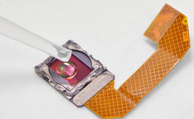
“Imaging can be done directly on image sensor chips [after removal of the optics], a technique possible to exploit commercially for the development of inexpensive ‘mini microscopes’,” the researchers said. “Images can be transferred for analysis both visually and by computer vision both at point of care and at remote locations.”
The researchers used their mini microscopes to image the eggs of helminths (parasitic worms) present in the urine and stools of infected individuals. They first used this approach to detect urinary schistosomiasis, a severely under-diagnosed infection affecting hundreds of millions, primarily in sub-Saharan Africa.
For diagnostics at the point of care, they developed a highly specific pattern-recognition algorithm that analysed the image from the mini microscope and automatically detected the helminth eggs. Four out of five eggs observed visually could be identified.
The resolution of the microscope was dependent on the pixel size of the sensor but sufficient for identification of several pathogenic parasites. The researchers thus concluded that “parasitic worm eggs can be recognised by on-chip imaging using a webcam stripped of the optics” and that their method “offers both an inexpensive alternative to conventional microscopy and diagnostic assistance by computer vision”.
Method holds promise to make bioimaging X-ray machines smaller and more flexible
Researchers from Nanyang Technological University, Singapore have found a new way to produce...
A non-destructive way to locate microplastics in body tissue
Currently available analytical methods either destroy tissue in the body or do not allow...
Rapid imaging method shows how medicine moves beneath the skin
Researchers have developed a rapid imaging technique that allows them to visualise, within...


