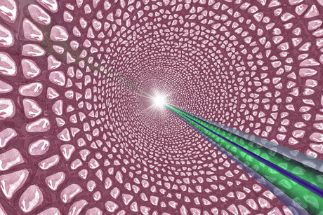
Probing cellular interactions

UTS Institute for Biomedical Materials & Devices (IBMD) new research offers the potential to see deeper into tissues to detect the biomarkers that are the very origins of disease.
“Traditional technologies can be invasive and require potentially toxic contrast agents or dyes,” said Dr Fan Wang from UTS (IBMD) and joint first author of the study published in Nature Communications.
“Another limitation is that you can’t use these technologies to find, or see into, individual diseased cells. You can’t achieve super-resolution imaging by penetrating ‘deep’ tissue. For example, with a CT scan you can see blood vessels but not the blood cells,” he said.
Dr Wang and the IBMD team instead use optical imaging techniques, photonics, allowing them to manipulate light at non-invasive lower wavelengths to take super-resolution imaging way beyond what is currently possible.
The UTS-led research demonstrates that optical techniques can operate at a scale several thousand times smaller than a cancer cell. Using a new nanomaterial, upconversion nanoparticles, or UCNPs, to probe thick tissue the researchers overcome two longstanding challenges to using photonics in super-resolution imaging — using low laser power to break through the optical diffraction limit and developing an efficient way to carry light beyond the first layers of human tissue to a depth that will allow single molecules, or nanomedicines, to be observed in 3D culture.
“Light scatters, the deeper you penetrate tissue with light the more it is absorbed and the more it scatters, impairing the resolution,” said Dr Fan Wang.
“We demonstrated that there is an optical narrow window in the near infrared (NIR) wavelength range, a sweet spot if you like, that allows light to penetrate deeper into tissue, beyond 50 nanometres, which, up until now, has been considered the limit of optical detection,” he said.
Having overcome this first fundamental challenge the team then used the unique emission wavelengths of a special type of UCNP (upconversion super dots) in the NIR range to act as a probe.
“Having broken through the 50 nm limit, we demonstrated the technology at a liver, brain or kidney tissue depth of 100 microns [100,000 nm] where you could still see a single molecule, which means there is the potential to detect a single disease biomarker. This is a very powerful tool for nanomedicine,“ Dr Wang said.
Professor Shaun Jackson, Director of the Heart Research Institute at University of Sydney, and co-author on the paper, said this proof-of-concept research is a significant advancement in a field that is becoming increasingly sophisticated for clinical researchers.
“This group is one of the few groups in the world doing this type of research. The prospect of being able to observe the physiological and pathological processes of disease in human tissue and then take those insights into clinical research is very exciting,” he said.
“In the future the hope is that the technology can be miniaturised and used, for example, in endoscopic devices to get clearer diagnoses,” he said.
UTS IBMD PhD candidate, and joint first author, Chaohao Chen said that this type of nanoscopy (microscopy at the nanoscale) has a number of advantages that will make it easier to commercialise than other super-resolution imaging technologies.
“It is easy to operate and very stable, which means the system doesn’t need a lot of servicing to keep it operating properly. The result is a technology that can be taken from the laboratory to industry at a relatively low cost,” he said.
IBMD Director Professor Dayong Jin said that this is exactly the type of technology that the institute was set up to achieve and follows on from the research for which he was awarded the 2017 Prime Ministers Prize for Science.
“This is our core business, fundamental science exploration in order to translate, transform photonics and material technology into disruptive biomedical devices and technology that everyone can access.
“We have answered the questions — yes, you can use optical methods for deep tissue penetration; yes, you can use optical methods for super-resolution imaging. Engineers can take this to the next level,” he said.
Future research will explore live cell super-resolution imaging to probe cellular interactions and observe in real time how nanomedicines are transported inside cells.
The Institute for Biomedical Materials & Devices (IBMD) at UTS Faculty of Science is transforming advances in photonics and materials into revolutionary biomedical technologies. Working closely with researchers and R&D partners around the globe, IBMD is delivering interdisciplinary research in nanophotonics, nanomaterials, biomaterials engineering, point-of-care diagnostics technologies and super-resolution bio-imaging.
A non-destructive way to locate microplastics in body tissue
Currently available analytical methods either destroy tissue in the body or do not allow...
Rapid imaging method shows how medicine moves beneath the skin
Researchers have developed a rapid imaging technique that allows them to visualise, within...
Fluorescent molecules glow in water, enhancing cell imaging
Researchers have developed a new family of fluorescent molecules that glow in a surprising way,...


