
Biosensors open a portal to improving cancer treatment
UK and Australian scientists are using tiny biosensors and fluorescence resonance energy transfer (FRET) imaging techniques to visualise how cancers spread and how active cancer cells respond to a particular drug.
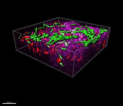
By visualising cancer cells in real time and in three dimensions, these technologies enable the researchers to pinpoint regions of poor drug delivery deep within a tumour. This enables drug delivery to be improved, which, in turn, improves clinical outcome for patients.
The study was performed by Dr Paul Timpson of the Garvan Institute of Medical Research and Professor Kurt Anderson of the Beatson Institute for Cancer Research in Glasgow, UK. PhD student Max Nobis studied the signalling protein ‘Src’, which becomes activated and drives invasive pancreatic cancer. He looked at how Src could best be deactivated by the small molecule inhibitor dasatinib, which is currently in phase II clinical trials.
“We have already shown that Src is activated in pancreatic tumours and we knew that dasatinib deactivates Src and could partially reduce the spread of this form of cancer. Through a collaborative partner in the US, we had access to FRET imaging technology,” said Timpson.
“Until now, we have been limited to studying tumour signalling in two dimensions - and lacked a dynamic way of reporting on drug targeting in live tumour tissue. Nanotechnology opens up a portal into living tissue that allows us to watch cancers spreading, and to determine which parts of a tumour we should be targeting with drugs.”
These techniques can also reveal how much, how often and how long to administer drugs. In addition, using these techniques in preclinical models of the disease, researchers can guide the use of combination therapies and enhance drug delivery by breaking up the tissue surrounding a tumour.
It has been hard to treat pancreatic tumours because they are extremely dense with collagen and have poor blood vessel networks for delivering drugs.
Anderson observed that combination therapies can be used to break down collagen, weakening tumour architecture and making it easier to get the drugs where they need to be.
“The trick is to break down the structure just enough to get the drug in, but not so much that you damage the organ itself,” he said. “These new FRET technologies help us gauge what is just enough and not too much.”
The findings were published in the journal Cancer Research.
Australia's first cases of H5 avian influenza confirmed
Positive results for H5 avian influenza (bird flu) have been confirmed in two seabirds found in...
AusBiotech partners with Tenmile
Designed to support Australia's homegrown life sciences innovation, AusBiotech has announced...
Australian CDC issues update in wake of Ebola outbreak
After the WHO determined the outbreak of Ebola in the DRC and Uganda to be a public health...


