
Scorpions and spectrometers to improve cancer surgery
Wednesday, 23 October, 2013
Scorpions could hold the secret to better cancer treatment, according to researchers from the Queensland Tropical Health Alliance (QTHA). The alliance recently acquired a $1.2 million nuclear magnetic resonance (NMR) spectrometer and facility to investigate the structure of promising molecules, some of which are found in scorpion venom.
Professor Norelle Daly’s research into scorpion venom began as a collaboration with Professor David Craik at the University of Queensland (UQ) and Dr Jim Olson at the Fred Hutchinson Cancer Research Center in Seattle. She explained, “The scorpion toxin, chlorotoxin, has been shown to bind to tumour cells. Therefore, labelling the toxin with a dye allows the tumour cells to be visualised.”
Such visualisation would help to more clearly distinguish the margin between cancerous and healthy cells during surgery, Professor Daly said. She hopes to find features in the scorpion toxin which “might one day help surgeons more accurately target cancers, including brain tumours”.

After applying for an Australian Research Council Future Fellowship to join the QTHA at James Cook University (JCU), she learned that JCU’s Professor Alex Loukas and Dr Jason Mulvenna, who had recently established laboratories with high-end mass spectrometry equipment, were also keen to establish an NMR facility.
“As my research requires an NMR, we put in an application for a LIEF grant from the Australian Research Council to partially fund an NMR spectrometer,” Professor Daly said.
The researchers were awarded $630,000 to support the high-resolution, high-throughput NMR facility, while the rest of the money was put in by JCU. Dr David Wilson was also funded by JCU to manage the facility.
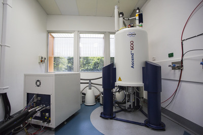
Dr Wilson explained that NMR occurs when the nuclei of certain atoms, placed in magnetic fields, absorb and re-emit electromagnetic radiation. “Some nuclei experience this phenomenon and others do not,” he said, “so we can measure this to try and understand the structure of these molecules, including those in scorpion venom.”
The facility’s NMR spectrometers, featuring cryogenically cooled probes, are also being used to study the structures of small proteins and small molecules isolated from creatures such as spiders, cone snails, jellyfish and hookworms, as “the structural information gained from these studies might be useful in the drug design process”, Professor Daly said.
She further noted that NMR spectroscopy is “highly suited to studying the [3D] structures of small proteins that are often present in venomous creatures”, as it is one of only two techniques which can reveal a high level of structural detail (the second being X-ray crystallography).
Meanwhile, outside the facility the original collaboration continues. Professor Daly and her research colleagues are doing some of the characterisation studies together, while Dr Olson is exploring whether tumour imaging can be used to help surgeons more effectively remove tumours.
Prototype promises to build light analysis directly into imaging hardware
Thanks to a new chip, researchers believe they may be able to help cameras capture details that...
Ultrasound improves protein recovery from cauliflower waste
Abundant but often discarded during processing, cauliflower leaves contain protein and dietary...
Can yawning advance the study of neurodegenerative diseases?
Australian neuroscientists using MRI scans to monitor flow of cerebrospinal fluid during yawning...


