
Technological breakthrough for skin regeneration
Researchers from the Beckman Institute for Advanced Science and Technology at the University of Illinois at Urbana-Champaign have demonstrated new multimodal optical microscopy technology which, when coupled with advanced image co-registration algorithms, can account for soft-tissue deformations that may occur over a course of weeks and months. Their report has been published in the journal Technology.
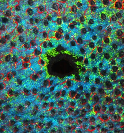
The technology can visualise cellular-level structural, functional and biomechanical data and provide new insights into complex processes in living tissue, such as skin. The technology not only offers a new tool for basic science and preclinical investigations, but also offers the potential for human clinical investigations into the dynamics of skin regeneration and repair, the incorporation of engineered skin constructs, the effectiveness of new therapies for treating skin conditions such as non-healing diabetic ulcers and for skin cosmesis applications.
“This is a remarkable combination of optical hardware and software technology that provides not only beautiful and fascinating images of dynamic microscopic processes, but also opens the door for future clinical applications,” said Dr Stephen Boppart, a senior author on the paper. “Multimodal imaging, and the ability to track cell and tissue dynamics over many months, will yield enormous volumes of data that will undoubtedly provide clues into many complex processes in skin.”
By spatially co-registering images from multiple modalities including optical coherence, two-photon excitation, second harmonic and phase-variance microscopy, as well as co-registering these volumetric image datasets temporally at timepoints that span several months, the investigators were able to visualise processes in wound healing and skin regeneration, as well as angiogenesis and tissue biomechanics including skin contraction.
“This is a nice example of where advances in imaging technology can enable both biological discovery as well as clinical applications,” said Dr Ben Graf, the lead author on the paper.
The team now plans to translate these technologies into clinical applications, such as investigating differences in cell and tissue dynamics over short- and long-term time periods for skin regeneration in normal and diabetic preclinical models. It is well known that diabetic patients suffer from chronic non-healing skin ulcers that frequently result in lower limb amputations. New therapies, including the use of stem cells, have shown promise, but the mechanisms for repair and regeneration are poorly understood.
With the large volumes of spatial and temporal data generated from this system, the investigators are also exploring how they can exploit the co-registered data to discover new biomarkers and diagnostic parameters that may provide clues into fundamental disease mechanisms.
The research was supported in part by grants from the National Science Foundation.
Prototype promises to build light analysis directly into imaging hardware
Thanks to a new chip, researchers believe they may be able to help cameras capture details that...
Ultrasound improves protein recovery from cauliflower waste
Abundant but often discarded during processing, cauliflower leaves contain protein and dietary...
Can yawning advance the study of neurodegenerative diseases?
Australian neuroscientists using MRI scans to monitor flow of cerebrospinal fluid during yawning...


